A copy of this work was available on the public web and has been preserved in the Wayback Machine. The capture dates from 2019; you can also visit the original URL.
The file type is application/pdf.
Filters
Multimodal Correlative Preclinical Whole Body Imaging and Segmentation
2016
Scientific Reports
Preserved Fulltext
This paper presents a novel approach for whole body segmentation of small animals in a multimodal setting of MR, CT and optical imaging. ...
Segmentation of anatomical tissues is fundamental for accurate and robust multi-modal correlative imaging and quantitative analysis in preclinical research. ...
Acknowledgements The authors would like to thank Nava Nevo for her help in tumor injection and advice on tumor delineation. ...
doi:10.1038/srep27940
pmid:27325178
pmcid:PMC4914843
fatcat:y3z32ebeonegxaa6fy4mkdaw2a
Tumor Sensitive Matching Flow: An Approach for Ovarian Cancer Metastasis Detection and Segmentation
[chapter]
2012
Lecture Notes in Computer Science
Preserved Fulltext
A copy of this work was available on the public web and has been preserved in the Wayback Machine. The capture dates from 2019; you can also visit the original URL.
The file type is application/pdf.
The routine machine learning strategies to locate ovarian tumors work poorly because the tumors spread randomly to the entire abdomen. ...
The proposed algorithm was validated on contrast-enhanced CT data from 11 patients with 26 metastases. 84.6% of metastases were successfully detected, and false positive per patient was 1.2. ...
Elise Kohn for helpful comments. ...
doi:10.1007/978-3-642-33612-6_20
fatcat:ml7lwlh5lzd6rj7qacvbynrvye
Medical Images Segmentation Based on Unsupervised Algorithms: A Review
2021
Qubahan Academic Journal
Preserved Fulltext
A copy of this work was available on the public web and has been preserved in the Wayback Machine. The capture dates from 2021; you can also visit the original URL.
The file type is application/pdf.
The medical image is divided into regions based on the specific descriptions, such as tissue/organ division in medical applications for border detection, tumor detection/segmentation, and comprehensive ...
(Magnetic Resonance Imaging), So segmentation of medical images is considered one of the most important medical imaging processes because it extracts the field of interest from the Return on investment ...
MRI images & CT-scan K-mean clustering algorithm Relative differences between (0.63-1.75) percent for MRI images and (0.34-1.51 percent) for CT images and measured surface areas for divided tumor areas ...
doi:10.48161/qaj.v1n2a51
fatcat:nc63bvlkdjewphs5yb2blk4qeq
Classification and Stage Prediction of Lung Cancer using Convolutional Neural Networks
2019
VOLUME-8 ISSUE-10, AUGUST 2019, REGULAR ISSUE
Preserved Fulltext
A copy of this work was available on the public web and has been preserved in the Wayback Machine. The capture dates from 2022; you can also visit the original URL.
The file type is application/pdf.
The CT scanned lung images should be involved in image classification processing for earlier prediction of stages and treatment diagnosis. ...
The extracted fine-grained training data through deep learning are utilized for the classification using Convolution Neural Network (CNN). ...
Pseudo code for this proposed model: Step 1: Obtain the input CT lung image from the user Step 2: Preprocess the image to get the appropriate scale 500 x 500 pixels This model can be experimentally verified ...
doi:10.35940/ijitee.j9146.0881019
fatcat:ums4iu3vwzfqxlzmugjcq2yvwe
Deep Learning in Breast Cancer Imaging: A Decade of Progress and Future Directions
[article]
2024
arXiv
pre-print
Preserved Fulltext

A copy of this work was available on the public web and has been preserved in the Wayback Machine. The capture dates from 2023; you can also visit the original URL.
The file type is application/pdf.
Note that this fulltext copy is not of the "primary" version of this work. The version it corresponds to is:
2023 pre-print arXiv:2304.06662v3 fatcat:w2bqc2lqunhrfmx456tjqnxguqOther Versions
Drawn from the findings of this survey, we present a comprehensive discussion of the challenges and potential avenues for future research in deep learning-based breast cancer imaging. ...
This paper provides an extensive review of deep learning-based breast cancer imaging research, covering studies on mammogram, ultrasound, magnetic resonance imaging, and digital pathology images over the ...
., 2022) for tumor segmentation + lightweight multi-scale network for classification + iterative feature refinement.Zhai et al., 2022). ...
arXiv:2304.06662v4
fatcat:t5nvpybawjhfhiw4h2bekozo74
Front Matter: Volume 9788
2016
Medical Imaging 2016: Biomedical Applications in Molecular, Structural, and Functional Imaging
Preserved Fulltext

A copy of this work was available on the public web and has been preserved in the Wayback Machine. The capture dates from 2018; you can also visit the original URL.
The file type is application/pdf.
The publisher is not responsible for the validity of the information or for any outcomes resulting from reliance thereon. ...
Please use the following format to cite material from these proceedings: Publication of record for individual papers is online in the SPIE Digital Library. ...
of the spine from IVDs and
vertebra segmentations [9788-65]
9788 1V
Three modality image registration of brain SPECT/CT and MR images for quantitative
analysis of dopamine transporter imaging [9788 ...
doi:10.1117/12.2240426
dblp:conf/mibam/X16
fatcat:msxwbg54kbegriwgvgcw4dnzu4
A Cascaded Feature Extraction for Diagnosis of Ovarian Cancer in CT Images
2022
International Journal of Advanced Computer Science and Applications
Preserved Fulltext
A copy of this work was available on the public web and has been preserved in the Wayback Machine. The capture dates from 2023; you can also visit the original URL.
The file type is application/pdf.
To improve the feature extraction and reduce the error, use a cascading technique for the feature extraction. RSO also helps to efficiently optimize the DCNN features from the images. ...
This paper proposed ovarian cancer detection in the ovarian image using joint feature extraction and an efficient Net model. ...
To capture the prognostic biomarkers (DL feature) of HGSOC, 8917 CT images from the feature learning cohort were used to train a unique DL network. ...
doi:10.14569/ijacsa.2022.0131235
fatcat:qg777ll345ck5fyantk5eetfhq
ECPC-IDS:A benchmark endometrail cancer PET/CT image dataset for evaluation of semantic segmentation and detection of hypermetabolic regions
[article]
2023
arXiv
pre-print
Preserved Fulltext
A copy of this work was available on the public web and has been preserved in the Wayback Machine. The capture dates from 2024; you can also visit the original URL.
The file type is application/pdf.
Note that this fulltext copy is not of the "primary" version of this work. The version it corresponds to is:
2023 pre-print arXiv:2308.08313v1 fatcat:xmss2ufxzvgutlhbbqnni5oe4mOther Versions
PET/CT Image Dataset for Evaluation of Semantic Segmentation and Detection of Hypermetabolic Regions (ECPC-IDS) are published. ...
Specifically, the segmentation section includes PET and CT images, with a total of 7159 images in multiple formats. ...
Yingying Hou from Foreign Studies College in Northeastern University, China, for her professional English proofreading in this paper. ...
arXiv:2308.08313v3
fatcat:awl7fcmlp5fetlnez2zfh3ud2m
Liver Tumors Segmentation Using 3D SegNet Deep Learning Approach
2023
Computer systems science and engineering
Preserved Fulltext
A copy of this work was available on the public web and has been preserved in the Wayback Machine. The capture dates from 2022; you can also visit the original URL.
The file type is application/pdf.
A standard data set was used to test the proposed model for liver CT scans and the tumor accuracy in the training phase. ...
In this article, a Deep Learning (DL) model is implemented and modified to fit liver CT segmentation, and a semantic pixel classification of road scenes is recommended. ...
Segmenting 69 CT (2D) images was the first step in the proposed method. All CT images came from the liver imaging Atlas (an online reference for liver imaging). ...
doi:10.32604/csse.2023.030697
fatcat:vjmhp2viyvdudc2kb25h2r2oty
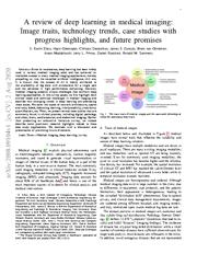
A review of deep learning in medical imaging: Image traits, technology trends, case studies with progress highlights, and future promises
[article]
2020
arXiv
pre-print
Preserved Fulltext
A copy of this work was available on the public web and has been preserved in the Wayback Machine. The capture dates from 2020; you can also visit the original URL.
The file type is application/pdf.
Other Versions
However, medical imaging presents unique challenges that confront deep learning approaches. ...
Since its renaissance, deep learning has been widely used in various medical imaging tasks and has achieved remarkable success in many medical imaging applications, thereby propelling us into the so-called ...
For multi-organ segmentation, Shi et al. ...
arXiv:2008.09104v1
fatcat:z2gic7or4vgnnfcf4joimjha7i
Machine Learning Methods for Histopathological Image Analysis: A Review
[article]
2021
arXiv
pre-print
Preserved Fulltext
A copy of this work was available on the public web and has been preserved in the Wayback Machine. The capture dates from 2021; you can also visit the original URL.
The file type is application/pdf.
Histopathological images (HIs) are the gold standard for evaluating some types of tumors for cancer diagnosis. ...
In this paper, we present a review on machine learning methods for histopathological image analysis, including shallow and deep learning methods. ...
Afterwards, ensembles of CNNs were trained to extract multi-context information from multi-scale images. The latter stage extracted both global and local features of breast cancer tumors. George et al ...
arXiv:2102.03889v1
fatcat:ylrsildl4nenho22erndvpjcjy
AMIGO: Sparse Multi-Modal Graph Transformer with Shared-Context Processing for Representation Learning of Giga-pixel Images
[article]
2023
arXiv
pre-print
Preserved Fulltext
A copy of this work was available on the public web and has been preserved in the Wayback Machine. The capture dates from 2023; you can also visit the original URL.
The file type is application/pdf.
Note that this fulltext copy is not of the "primary" version of this work. The version it corresponds to is:
2023 pre-print arXiv:2303.00865v1 fatcat:6oct37isnjd2rm7sk4zfa52pim
Multiple instance learning (MIL) has become the conventional approach to process WSIs, in which these images are split into smaller patches for further processing. ...
In this paper, by defining the novel concept of shared-context processing, we designed a multi-modal Graph Transformer (AMIGO) that uses the celluar graph within the tissue to provide a single representation ...
image types (e.g., histopathology images, CT scans, and MRI scans) and numerous tasks (e.g., classification, segmentation, and survival prediction) [6, 11, 27, 29, 35, 37, 43] . ...
arXiv:2303.00865v2
fatcat:ldu663pmrvcclaga4zfa3ivhlu
2020 Index IEEE Journal of Biomedical and Health Informatics Vol. 24
2020
IEEE journal of biomedical and health informatics
Preserved Fulltext
A copy of this work was available on the public web and has been preserved in the Wayback Machine. The capture dates from 2021; you can also visit the original URL.
The file type is application/pdf.
., A Globalized Model for Mapping Wearable Seismocardiogram Signals to Whole-Body Ballistocardiogram Signals Based on Deep Learning; JBHI May 2020 1296-1309 Herskovic, V., see Saint-Pierre, C., JBHI Jan ...
., +, JBHI Jan. 2020
14-16
M 3 Lung-Sys: A Deep Learning System for Multi-Class Lung Pneumonia
Screening From CT Imaging. ...
., +, JBHI Dec. 2020
3595-3605
M 3 Lung-Sys: A Deep Learning System for Multi-Class Lung Pneumonia
Screening From CT Imaging. ...
doi:10.1109/jbhi.2020.3048808
fatcat:iifrkwtzazdmboabdqii7x5ukm
2021 Index IEEE/ACM Transactions on Computational Biology and Bioinformatics Vol. 18
2022
IEEE/ACM Transactions on Computational Biology & Bioinformatics
Preserved Fulltext
A copy of this work was available on the public web and has been preserved in the Wayback Machine. The capture dates from 2022; you can also visit the original URL.
The file type is application/pdf.
The Author Index contains the primary entry for each item, listed under the first author's name. ...
-that appeared in this periodical during 2021, and items from previous years that were commented upon or corrected in 2021. ...
., +, TCBB March-April 2021 562-574 + Check author entry for coauthors Biomedical imaging A Deep Segmentation Network of Multi-Scale Feature Fusion Based on Attention Mechanism for IVOCT Lumen Contour. ...
doi:10.1109/tcbb.2021.3136340
fatcat:bjvb334webfovh4nsc7oeds3di
Deep Interactive Segmentation of Medical Images: A Systematic Review and Taxonomy
[article]
2024
arXiv
pre-print
Preserved Fulltext

A copy of this work was available on the public web and has been preserved in the Wayback Machine. The capture dates from 2024; you can also visit the original URL.
The file type is application/pdf.
Other Versions
Interactive segmentation is a crucial research area in medical image analysis aiming to boost the efficiency of costly annotations by incorporating human feedback. ...
In recent years, deep learning-based approaches have propelled results to a new level causing a rapid growth in the field with 121 methods proposed in the medical imaging domain alone. ...
head-and-neck cancer PET/CT/MRI Link Zhuang et al. 2 [90] Scribbles Exponential Geodesic Maps Train Active Learning brain tumors, liver tumors CT, MRI Link GtG [91] Clicks Gaussian Heatmaps Sim Iter Distance ...
arXiv:2311.13964v2
fatcat:bzxla3flzna5ze7nmm5nq33rju
« Previous
Showing results 1 — 15 out of 493 results