A copy of this work was available on the public web and has been preserved in the Wayback Machine. The capture dates from 2019; you can also visit the original URL.
The file type is application/pdf.
Filters
Resection-induced brain-shift compensation using vessel-based methods
2018
Medical Imaging 2018: Image-Guided Procedures, Robotic Interventions, and Modeling
Preserved Fulltext
Most brain-shift compensation methods address the problem of updating preoperative images to reflect brain deformations following the craniotomy and dura opening. ...
While more patients must be included, these preliminary results show that vesselbased methods are well suited to compensate for resection-induced brain-shift, but better outcomes in complex cases still ...
Figure 3 . 3 Brains-shift compensation for case 4, resection of a deep tumor. An anatomical landmark of the Sylvian sulcus is pointed on the pMR, iUS and updated pMR images. ...
doi:10.1117/12.2293640
dblp:conf/miigp/MorinCRLPPC18
fatcat:qcgtkvbz5vhg3faodooppltkea
Estimation of Brain Deformation for Volumetric Image Updating in Protoporphyrin IX Fluorescence-Guided Resection
2010
Stereotactic and Functional Neurosurgery
Preserved Fulltext
A copy of this work was available on the public web and has been preserved in the Wayback Machine. The capture dates from 2019; you can also visit the original URL.
The file type is application/pdf.
Conclusion: Implementation of brain deformation modeling in FGR shows promise for increasing the accuracy of neurosurgical guidance in the delineation and resection of brain tumors. ...
sound data was used to generate updated MR images. ...
Roberts serves on the data monitoring committee for a Medtronic deep brain stimulation study. ...
doi:10.1159/000258143
pmid:19907205
pmcid:PMC2813794
fatcat:epjqftukjrcibp3osceseqdxry
Framework and Bio-Mechanical Model for a Per-Operative Image-Guided Neuronavigator Including 'Brain-Shift' Compensation
[article]
2006
arXiv
pre-print
Preserved Fulltext
The Internet Archive has a preservation copy of this work in our general collections.
The file type is application/pdf.
In this paper we present a methodology to adress the problem of brain tissue deformation referred to as "brainshift". ...
as surgical procedures that modify the brain structure, like tumour or tissue resection. ...
Figure 3 -F: Gravity, cyst drainage and tissue resection cause the brain to sag.
CONCLUSION We proposed a general framework for a image-guided model-updated neuronavigator for tumor resection. ...
arXiv:physics/0610185v1
fatcat:53zvuppmujah5hsd25ioxa6bhi
Modeling of Retraction and Resection for Intraoperative Updating of Images
2001
Neurosurgery
Preserved Fulltext
A copy of this work was available on the public web and has been preserved in the Wayback Machine. The capture dates from 2004; you can also visit the original URL.
The file type is application/pdf.
CONCLUSION: The results presented demonstrate that complex surgical events such as tissue retraction and resection can be incorporated intraoperatively into the model-updating process for brain shift compensation ...
RESULTS: Retraction and resection techniques are demonstrated to accurately reflect intraoperative events, thus providing an approach for near-real-time image-updating in the OR. ...
AnalyzeAVW software was provided in collaboration with the Mayo Foundation. ...
doi:10.1097/00006123-200107000-00012
pmid:11440463
fatcat:yvcyeva6t5fejhceut33yiwdju
Modeling of Retraction and Resection for Intraoperative Updating of Images
2001
Neurosurgery
Preserved Fulltext
A copy of this work was available on the public web and has been preserved in the Wayback Machine. The capture dates from 2004; you can also visit the original URL.
The file type is application/pdf.
CONCLUSION: The results presented demonstrate that complex surgical events such as tissue retraction and resection can be incorporated intraoperatively into the model-updating process for brain shift compensation ...
RESULTS: Retraction and resection techniques are demonstrated to accurately reflect intraoperative events, thus providing an approach for near-real-time image-updating in the OR. ...
AnalyzeAVW software was provided in collaboration with the Mayo Foundation. ...
doi:10.1227/00006123-200107000-00012
fatcat:plyk45pgcbgktng3rn7ubaxm7a
Intraoperative Imaging Modalities and Compensation for Brain Shift in Tumor Resection Surgery
2017
International Journal of Biomedical Imaging
Preserved Fulltext
A copy of this work was available on the public web and has been preserved in the Wayback Machine. The capture dates from 2019; you can also visit the original URL.
The file type is application/pdf.
Clinical experience of using intraoperative imaging modalities, details about registration, and modeling methods in connection with brain shift in tumor resection surgery are the focuses of this review ...
The purpose of this paper is to present a review of publications concerning different aspects of intraoperative brain shift especially in a tumor resection surgery such as intraoperative imaging systems ...
Compensation Methods Guided by Intraoperative Image Data One strategy to compensate for intraoperative brain shift in tumor resection surgery is to register intraoperative two-or three-dimensional image ...
doi:10.1155/2017/6028645
pmid:28676821
pmcid:PMC5476838
fatcat:hb3tljwacnc2po4dbmibs27x3e
Framework for a Low-Cost Intra-Operative Image-Guided Neuronavigator Including Brain Shift Compensation
2007
IEEE Engineering in Medicine and Biology Society. Conference Proceedings
Preserved Fulltext
A copy of this work was available on the public web and has been preserved in the Wayback Machine. The capture dates from 2017; you can also visit the original URL.
The file type is application/pdf.
In this paper we present a methodology to address the problem of brain tissue deformation referred to as 'brain-shift'. ...
model which can take into account tissue deformations and surgical procedures that modify the brain structure, like tumour or tissue resection. ...
neurosurgical systems a module to compensate brain deformations by updating the pre-operative images and planning according to intra-operative brain shape changes. ...
doi:10.1109/iembs.2007.4352429
pmid:18002095
fatcat:pjsk6a3jujcbziqjxkspdcoyfq
Bio-mechanical model of the brain for a per-operative image-guided neuronavigator compensating for "brain-shift" deformations
2007
Computer Methods in Biomechanics and Biomedical Engineering
Preserved Fulltext
A copy of this work was available on the public web and has been preserved in the Wayback Machine. The capture dates from 2017; you can also visit the original URL.
The file type is application/pdf.
In order to face this problem, authors have proposed to add to actual image guided neurosurgical systems a module to compensate brain deformations by updating the preoperative images and planning according ...
Hastreiter et al. [1] observed a great variability of the brain-shift ranging up to 24 mm for cortical displacement and exceeding 3 mm for the deep tumor margin; the authors claim for a non-correlation ...
The resection update of the system takes about 5 to 10 seconds on the aforementioned mesh. ...
doi:10.1080/10255840701479057
fatcat:i4p2zi6cjjazxkm67bv7lgiffu
A Fast and Efficient Method to Compensate for Brain Shift for Tumor Resection Therapies Measured Between Preoperative and Postoperative Tomograms
2010
IEEE Transactions on Biomedical Engineering
Preserved Fulltext
A copy of this work was available on the public web and has been preserved in the Wayback Machine. The capture dates from 2010; you can also visit the original URL.
The file type is application/pdf.
In this paper, an efficient paradigm is presented to correct for brain shift during tumor resection therapies. ...
Index Terms-Brain shift, finite elements, image deformation, image-guided surgery, inverse model. ...
ACKNOWLEDGMENT The authors would like to thank the Resident Surgeons, the OR Staff, and the Radiology Department at Vanderbilt University for their help in data collection. ...
doi:10.1109/tbme.2009.2039643
pmid:20172796
pmcid:PMC2891363
fatcat:szjjjbjdgnbhvkob4yzdfotoem
Intraoperative brain tumor resection cavity characterization with conoscopic holography
2012
Medical Imaging 2012: Image-Guided Procedures, Robotic Interventions, and Modeling
Preserved Fulltext
A copy of this work was available on the public web and has been preserved in the Wayback Machine. The capture dates from 2017; you can also visit the original URL.
The file type is application/pdf.
improve the accuracy of image-guided surgical interventions in the presence of soft tissue deformations. ...
Laser range scan, instrument swabbing, and conoscopic holography data sets were collected from two patients undergoing brain tumor resection therapy at Vanderbilt University Medical Center. ...
COMPENSATING FOR DEFORMATION WITH MATHEMATICAL MODELS In this section, we give an overview of our specific framework for compensation of deformation in the brain using mathematical models. ...
doi:10.1117/12.911926
dblp:conf/miigp/SimpsonBCPSTWM12
fatcat:6gtykmedince7pbosqngegzud4
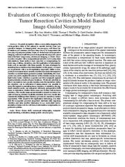
Evaluation of Conoscopic Holography for Estimating Tumor Resection Cavities in Model-Based Image-Guided Neurosurgery
2014
IEEE Transactions on Biomedical Engineering
Preserved Fulltext
A copy of this work was available on the public web and has been preserved in the Wayback Machine. The capture dates from 2017; you can also visit the original URL.
The file type is application/pdf.
for soft-tissue deformation compensation in guidance systems. ...
Some authors have reported on compensating for brain sag, swelling, retraction, and the application of pharmaceuticals such as mannitol with these models. ...
Dawant for helpful discussions on the nonrigid alignment of pre-and postoperative MRI neurosurgery data. This study would not have been ...
doi:10.1109/tbme.2014.2308299
pmid:24845293
pmcid:PMC4185972
fatcat:b2iszy456fejxe5gmnombwkzb4
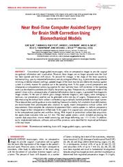
Near Real-Time Computer Assisted Surgery for Brain Shift Correction Using Biomechanical Models
2014
IEEE Journal of Translational Engineering in Health and Medicine
Preserved Fulltext
A copy of this work was available on the public web and has been preserved in the Wayback Machine. The capture dates from 2017; you can also visit the original URL.
The file type is application/pdf.
), updated images of the brain must be provided to the neuronavigation system in a timely manner for practical use in the operating room. ...
Once complete, the volumetric displacement field is used to update, i.e., deform, preoperative brain images to their intraoperative shifted state. ...
Benoit Dawant for providing the atlas-based brain segmentation code. ...
doi:10.1109/jtehm.2014.2327628
pmid:25914864
pmcid:PMC4405800
fatcat:mmiibms7hbdgfpz3wtvx6bo2qe
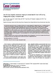
Use of virtual magnetic resonance imaging to compensate for brain shift during image-guided surgery: illustrative case
2022
Journal of Neurosurgery: Case Lessons
Preserved Fulltext
A copy of this work was available on the public web and has been preserved in the Wayback Machine. The capture dates from 2022; you can also visit the original URL.
The file type is application/pdf.
LESSONS EIF algorithms can be used with multimodal images (preoperative MRI and intraoperative CT) and create an updated virtual MRI data set to compensate for brain shift in neurosurgery and aid in maximum ...
OBSERVATIONS The authors described a case in which an image fusion algorithm was used in conjunction with an intraoperative computed tomography (CT) system to compensate for brain shift during resection ...
An updated virtual iMRI data set was generated and used to compensate for brain shift and aid in complete resection of the brainstem tumor. ...
doi:10.3171/case21683
pmid:35733635
pmcid:PMC9204912
fatcat:u36qcdgkhrcdfgikoukm7lwqgu
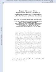
Doppler Ultrasound Driven Biomechanical Model of the Brain for Intraoperative Brain-Shift Compensation: A Proof of Concept in Clinical Conditions
[chapter]
2012
Studies in Mechanobiology, Tissue Engineering and Biomaterials
Preserved Fulltext
A copy of this work was available on the public web and has been preserved in the Wayback Machine. The capture dates from 2012; you can also visit the original URL.
The file type is application/pdf.
Yet, image-guided neurosurgery faces an important issue for large skull openings where brain soft-tissues can exhibit large deformations in the course of surgery. ...
The system, tested on a patient presenting a large meningioma, was able to compensate within seconds for the intraoperatively observed brainshift, reducing the mean error on tumor margin localization from ...
Gilles Francony from the surgical reanimation unit (URC) at the Hospital Michallon for their advice and help during this study. hal-00706803, version 1 -11 Jun 2012 ...
doi:10.1007/8415_2012_119
fatcat:id7vypjyyjbzrk2q4h54txxftq
Intraoperative Brain Shift Compensation Using a Hybrid Mixture Model
[chapter]
2018
Lecture Notes in Computer Science
Preserved Fulltext
A copy of this work was available on the public web and has been preserved in the Wayback Machine. The capture dates from 2019; you can also visit the original URL.
The file type is application/pdf.
Brain deformation (or brain shift) during neurosurgical procedures such as tumor resection has a significant impact on the accuracy of neuronavigation systems. ...
Compensating for this deformation during surgery is essential for effective guidance. ...
[5] employed B-spline based elastic image registration to compensate for brain shift, using pre-and intraoperative CBCT images (although, not during surgical tumor resection). ...
doi:10.1007/978-3-030-00937-3_14
fatcat:7e4to4arybhc5ncuad4aqdeneu
« Previous
Showing results 1 — 15 out of 1,019 results