A copy of this work was available on the public web and has been preserved in the Wayback Machine. The capture dates from 2006; you can also visit the original URL.
The file type is application/pdf.
Filters
Displacement estimation with co-registered ultrasound for image guided neurosurgery: A quantitative in vivo porcine study
2003
IEEE Transactions on Medical Imaging
Preserved Fulltext

For one of the animals studied, the US data was used in conjunction with a biomechanical model to nonrigidly re-register a baseline CT to the deformed brain. ...
Index Terms-Brain modeling, brain shift, image-guided neurosurgery, image registration, intraoperative ultrasound. ...
Extracting Bead Displacement Data In order to compare displacement measurements from CT and US data, the beads from all US and CT scans must be extracted and then mapped to the baseline CT volume using ...
doi:10.1109/tmi.2003.819293
pmid:14606670
fatcat:bqamhhddgnf3hpt53sag75rxxq
Verification of fetal brain responses by coregistration of fetal ultrasound and fetal magnetoencephalography data
2010
NeuroImage
Preserved Fulltext

A copy of this work was available on the public web and has been preserved in the Wayback Machine. The capture dates from 2020; you can also visit the original URL.
The file type is application/pdf.
For this aim either auditory or visual stimuli are presented and evoked brain activity or spontaneous activity is measured at the sensor level. ...
In the present report we overcome this limitation by a coregistration of volumetric ultrasound images with fMEG data. ...
For the extraction of the sphere from the US data, multiple points on the reconstructed US images are marked to determine the fetal head diameter and the center of the sphere. ...
doi:10.1016/j.neuroimage.2009.09.025
pmid:19778620
pmcid:PMC2948237
fatcat:7cfevyvrevhu5lddinio7ryxti
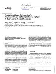
Estimation of Brain Deformation for Volumetric Image Updating in Protoporphyrin IX Fluorescence-Guided Resection
2010
Stereotactic and Functional Neurosurgery
Preserved Fulltext
A copy of this work was available on the public web and has been preserved in the Wayback Machine. The capture dates from 2019; you can also visit the original URL.
The file type is application/pdf.
Conclusion: Implementation of brain deformation modeling in FGR shows promise for increasing the accuracy of neurosurgical guidance in the delineation and resection of brain tumors. ...
sound data was used to generate updated MR images. ...
Roberts serves on the data monitoring committee for a Medtronic deep brain stimulation study. ...
doi:10.1159/000258143
pmid:19907205
pmcid:PMC2813794
fatcat:epjqftukjrcibp3osceseqdxry
Intraoperative image updating for brain shift following dural opening
2017
Journal of Neurosurgery
Preserved Fulltext

A copy of this work was available on the public web and has been preserved in the Wayback Machine. The capture dates from 2020; you can also visit the original URL.
The file type is application/pdf.
CONCLUSIONS-This study compensated for brain deformation caused by intraoperative dural opening using computational model-based assimilation of iSV cortical surface displacements. ...
Keywords image-guided neurosurgery; brain deformation; FEM model; sparse data; intraoperative stereovision; diagnostic and operative techniques Fan et al. ...
We acknowledge the support of Medtronic Navigation for use of the StealthStation S7 and Carl Zeiss (Carl Zeiss Surgical GmbH) for use of the OPMI Pentero operating microscope. ...
doi:10.3171/2016.6.jns152953
pmid:27611206
pmcid:PMC5549265
fatcat:v5wbnzytzjguth66jb3qkpfjr4
Graphical user interface for intraoperative neuroimage updating
2003
Medical Imaging 2003: Visualization, Image-Guided Procedures, and Display
Preserved Fulltext

A copy of this work was available on the public web and has been preserved in the Wayback Machine. The capture dates from 2005; you can also visit the original URL.
The file type is application/pdf.
Upon acquisition of registration data for patient position in the OR (using fiducial markers), the Matlab GUI displays ultrasound image overlays on patient specific, preoperative MR images. ...
Registration matrices are also applied to patientspecific anatomical models used for image updating. ...
model (FEM) to generate volumetric calculations of brain tissue displacement. ...
doi:10.1117/12.479761
dblp:conf/miigp/RickHRLSP03
fatcat:53bzbym345hgdeqbvh3jfbyh2m
A computational model for tracking subsurface tissue deformation during stereotactic neurosurgery
1999
IEEE Transactions on Biomedical Engineering
Preserved Fulltext

A copy of this work was available on the public web and has been preserved in the Wayback Machine. The capture dates from 2011; you can also visit the original URL.
The file type is application/pdf.
Specifically, we report on the initial development of a finite element model of brain tissue adapted from consolidation theory. ...
While the predicted tissue displacements differ from measured values by about 15%, they suggest that exploiting a physics-based computational framework for updating preoperative imaging databases during ...
For example, brain surface tracking devices could be used to extract boundary condition data for the model. ...
doi:10.1109/10.740884
pmid:9932343
fatcat:dhh7hn5mzbbizkvwaxl774ll2u
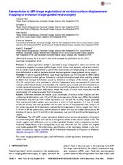
Stereovision to MR image registration for cortical surface displacement mapping to enhance image-guided neurosurgery
2014
Medical Physics (Lancaster)
Preserved Fulltext
A copy of this work was available on the public web and has been preserved in the Wayback Machine. The capture dates from 2020; you can also visit the original URL.
The file type is application/pdf.
The full three-dimensional (3D) displacement field was also extracted to drive a biomechanical brain deformation model, the results of which were reconciled with the reconstructed iSV surface as another ...
When the complete displacement map derived from surface registration was utilized, the resulting images generated from mechanical model updates were consistent in terms of both geometry (1-2 mm of model ...
As a second form of evaluation that invoked data from the entire coregistered image surface, we used the full surface displacement map generated by the surface registration technique as sparse data in ...
doi:10.1118/1.4894705
pmid:25281972
pmcid:PMC5176089
fatcat:af2j4rm3t5cynd7rudqtlckjey
FocalErrorNet: Uncertainty-aware focal modulation network for inter-modal registration error estimation in ultrasound-guided neurosurgery
[article]
2023
arXiv
pre-print
Preserved Fulltext

A copy of this work was available on the public web and has been preserved in the Wayback Machine. The capture dates from 2023; you can also visit the original URL.
The file type is application/pdf.
Intra-operative ultrasound (iUS) has been adopted to provide real-time images to track brain shift, and inter-modal (i.e., MRI-iUS) registration is often required to update the pre-surgical plan. ...
Therefore, we propose a novel deep learning technique based on 3D focal modulation in conjunction with uncertainty estimation to accurately assess MRI-iUS registration errors for brain tumor surgery. ...
Each coregistered iUS scan was deformed ten times. ...
arXiv:2307.14520v1
fatcat:na32gzmxrjbr5mnq2k5jwjwqym
Initial in-vivo analysis of 3D heterogeneous brain computations for model-updated image-guided neurosurgery
[chapter]
1998
Lecture Notes in Computer Science
Preserved Fulltext

A copy of this work was available on the public web and has been preserved in the Wayback Machine. The capture dates from 2018; you can also visit the original URL.
The file type is application/pdf.
Registration error resulting from intraoperative brain shift due to applied surgical loads has long been recognized as one of the most challenging problems in the field of frameless stereotactic neurosurgery ...
To address this problem, we have developed a 3-dimensional finite element model of the brain and have begun to quantify its predictive capability in an in vivo porcine model. ...
This data suggests that coregistered image studies performed preoperatively cannot accurately account for such shift which in turn could lead to surgical error when relied upon for navigational information ...
doi:10.1007/bfb0056261
pmid:26317118
pmcid:PMC4548975
fatcat:afips7pldrhfhox2btm57uglia
Image-guided neurosurgery at Brigham and Women's Hospital
2006
IEEE Engineering in Medicine and Biology Magazine
Preserved Fulltext

A copy of this work was available on the public web and has been preserved in the Wayback Machine. The capture dates from 2010; you can also visit the original URL.
The file type is application/pdf.
Acknowledgments The authors wish to thank Heather O'Leary, Pairash Saiviroonporn, Stephen Whalen, Jun Wada, and the Brain Science Foundation for their contributions as well as several NIH and NSF grants ...
The overall accuracy of the brain deformation is estimated based on edge features extracted from the images. ...
Recently, he has been studying the use of ultrasound for temporary disruption of the blood-brain barrier, which may allow for targeted drug delivery in the brain. ...
doi:10.1109/memb.2006.1705749
pmid:17020201
fatcat:pisbdm52irddbg5pkddgawmsie
SlicerDMRI: Diffusion MRI and Tractography Research Software for Brain Cancer Surgery Planning and Visualization
2020
JCO Clinical Cancer Informatics
Preserved Fulltext

A copy of this work was available on the public web and has been preserved in the Wayback Machine. The capture dates from 2020; you can also visit the original URL.
The file type is application/pdf.
In this article, we focus on a demonstration of SlicerDMRI as an informatics tool to enable end-to-end dMRI analyses in two retrospective imaging data sets from patients with high-grade glioma. ...
that surrounds brain tumors. ...
Intraoperative ultrasound data are overlaid on the presurgical MRI data for an integrated visualization. ...
doi:10.1200/cci.19.00141
pmid:32216636
pmcid:PMC7113081
fatcat:ixutwa2k65fpjhdgmm7smeffsu
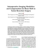
Intraoperative Imaging Modalities and Compensation for Brain Shift in Tumor Resection Surgery
2017
International Journal of Biomedical Imaging
Preserved Fulltext
A copy of this work was available on the public web and has been preserved in the Wayback Machine. The capture dates from 2019; you can also visit the original URL.
The file type is application/pdf.
Clinical experience of using intraoperative imaging modalities, details about registration, and modeling methods in connection with brain shift in tumor resection surgery are the focuses of this review ...
The consequences from the categorization and trends in the future are discussed at the end of this work. ...
The quantitative analyses show that coregistered iUS could measure the intraoperative displacement effectively and could be used with a computational model. ...
doi:10.1155/2017/6028645
pmid:28676821
pmcid:PMC5476838
fatcat:hb3tljwacnc2po4dbmibs27x3e
In vivo quantification of retraction deformation modeling for updated image-guidance during neurosurgery
2002
IEEE Transactions on Biomedical Engineering
Preserved Fulltext

A copy of this work was available on the public web and has been preserved in the Wayback Machine. The capture dates from 2007; you can also visit the original URL.
The file type is application/pdf.
We are developing the strategy of exploiting a computational model driven by sparse data obtained from intraoperative ultrasound and cortical surface tracking to warp preoperative images to reflect the ...
Overall, the level of quantitative agreement achieved in these experiments is encouraging for updating preoperative images to reflect tissue deformation resulting from retraction, especially since model ...
details from individual experiments which are important for highlighting the spatial characteristics of the model-data comparisons. ...
doi:10.1109/tbme.2002.800760
pmid:12148821
fatcat:hfwo2agplnbrhhzxpfg5vdhalm
Multi-modal learning-based pre-operative targeting in deep brain stimulation procedures
2016
2016 IEEE-EMBS International Conference on Biomedical and Health Informatics (BHI)
Preserved Fulltext

A copy of this work was available on the public web and has been preserved in the Wayback Machine. The capture dates from 2020; you can also visit the original URL.
The file type is application/pdf.
Deep brain stimulation, as a primary surgical treatment for various neurological disorders, involves implanting electrodes to stimulate target nuclei within millimeter accuracy. ...
Regression forests are used to learn a displacement vector of this point to the target. ...
To simplify model training, we define a bounding box that roughly covers the deep brain and constrain the samples to be uniformly drawn from this region. ...
doi:10.1109/bhi.2016.7455824
pmid:27754497
pmcid:PMC5042326
dblp:conf/bhi/LiuD16
fatcat:az6d6bwywjhufd7jq5fy6yz3a4
Ultrashort echo-time MRI versus CT for skull aberration correction in MR-guided transcranial focused ultrasound: In vitro comparison on human calvaria
2015
Medical Physics (Lancaster)
Preserved Fulltext

A copy of this work was available on the public web and has been preserved in the Wayback Machine. The capture dates from 2019; you can also visit the original URL.
The file type is application/pdf.
Purpose: Transcranial magnetic resonance-guided focused ultrasound (TcMRgFUS) brain treatment systems compensate for skull-induced beam aberrations by adjusting the phase and amplitude of individual ultrasound ...
Each target location was sonicated three times: once using aberration corrections calculated from the actual CT scan, once using corrections calculated from the MRI-derived virtual CT scan, and once without ...
Financial support was provided by the UVA Focused Ultrasound Center. G.W.M. receives research support from Siemens Healthcare. ...
doi:10.1118/1.4916656
pmid:25979016
fatcat:66nh43h2o5fsvlzgjdt47rd35i
« Previous
Showing results 1 — 15 out of 216 results